
Hitachi
Contact InformationContact Information

Healthcare Systems

Finance, Public Sector, Healthcare

    1. Establishment of Data Collection Platform for National Hospital Organization Clinical Data Archives

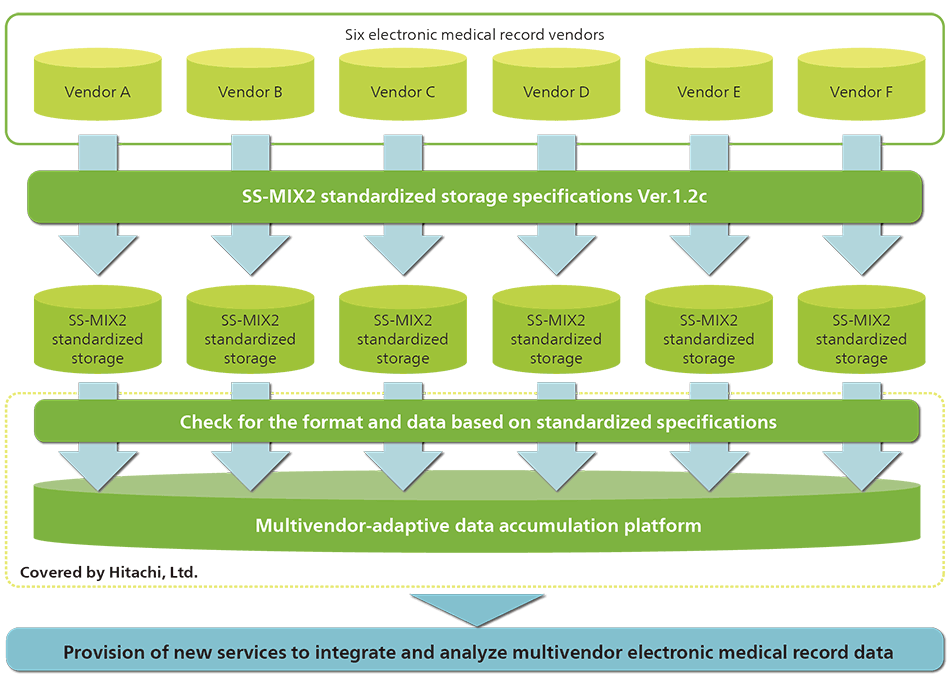
    In the business of building the National Hospital Organization Clinical Data Archives (NCDA) conducted by National Hospital Organization (NHO), Hitachi was in charge of constructing the data accumulation platform for the data center.

    NCDA collects medical data recorded in daily practice using the standardized structured medical information exchange 2 (SS-MIX2) standardized storage format, the standard specified by Ministry of Health, Labour and Welfare, without changing the electronic medical record system currently being used in the hospitals under the control of NHO. In addition, diagnosis procedure combination (DPC) evaluation data and medical expenses data accumulated separately will be integrated to create a database that will enable users to effectively analyze large amounts of complicated treatment information and will contribute to improving the quality of medical care and hospital management efficiency.

    By making use of the know-how fostered through building the NCDA, Hitachi aims to realize healthcare innovation by providing medical information utilization solutions.

    1. Outline of NCDA system

    2. Permanent Magnet Type 0.4T/0.3T Open MRI Renewed with the Newest Software

    Permanent magnet type 0.4 T and 0.3 T open magnetic resonance imaging (MRI) were updated with the newest software and usability was improved.

    The newly mounted auto slice positioning function is an original Hitachi function that can improve workflow by assisting users in setting the section to be imaged. When conducting a head examination, the imaging position is automatically set to the appropriate preset position and the burden on the operator can be reduced, which helps even inexperienced users to conduct an accurate inspection.

    In addition, Hitachi’s original parallel imaging technology, can reduce image deterioration caused by the patient’s motion. This function can be used in various types of images and any cross-section image, including T1/T2-weighted images and fluid attenuated inversion recovery (FLAIR). It can be effectively utilized in a clinical setting, since any body part can be imaged and any type of coil can be used.

    An open MRI has fewer units than a superconducting MRI, and is mainly composed of three units: a main unit gantry, a console, and a power supply system. What is more, it can be installed in a limited space since the leakage magnetic field is small, the size is compact, and no machine room is required. No water cooling system is needed, while it is necessary for a superconducting MRI.

    Hitachi will make use of its original technology and develop higher-value devices for users.

    2. Permanent magnet type open MRI 0.4 T (left), 0.3 T (right)

    3. Multi-slice CT Supria/Supria 64 with New Version 2.21

    3. Multi-slice CT Supria/Supria 64

    Supria, a 16-slice computed tomography (CT) scanner that was developed based on the concept of open and compact, and Supria 64*, a CT equipped with a 64-slice detector that makes it possible to take images at higher speed and in a broader range with the same sized gantry, are now compatible with the new version 2.21. This helps radiologists to read images and improve workflow, while power consumption is significantly lower than for existing CT scanners.

    In addition, the algorithm for iterative reconstruction was improved to reduce the image noise appearing when an examination is conducted with a lower level of X-rays, and it is equipped with rapid iterative reconstruction technology with high-speed image processing, which enables users to obtain images with the same quality as those taken using the conventional iterative reconstruction technology in about half the time. Images can be created immediately after CT scanning, which contributes to improved workflow.

    Eco mode has two functions to save energy. On-time standby reduces power consumption by appropriately controlling device operation, including the built-in cooling fans in the gantry. In addition, off-time mode can reduce standby power consumption by shortening the electric current time to the X-ray detector while CT scanner is not being used.

    *
    Supria 64 is the name for Supria with a 64-slice detector.

    4. Fetal Heart Rate Automatic Measurement Technology for Ultrasonic Diagnostic Equipment

    4. Examples of AutoFHR conducted in early pregnancy (9 weeks and 6 days, top) and in late pregnancy (32 weeks and 4 days, bottom)

    The fetal heart rate is one of the most basic and common measurement parameters when evaluating vitality, and it is also associated with a number of diseases. The fetal heart rate is generally measured using the Doppler method or M-mode method, although these measurement methods involve security concerns based on the principle of “as low as reasonably achievable” (ALARA), which recommends lower sound power as well as problems such as less objectivity due to the observation of a minute target of a few millimeters in size.

    The automated fetal heart rate (AutoFHR) measurement method automatically tracks fetal cardiac motion using B-mode imaging, and calculates the fetal heart rate. As a result, the issues associated with the traditional measurement methods have been resolved and fetal heart rate can be measured with high reproducibility. This technology is mounted on products in the ARIETTA series, Hitachi’s ultrasonic diagnostic equipment. It is expected that the measurement of the fetal heart rate in early pregnancy (6 weeks), which has not been carried out previously due to the difficulty with the procedure and safety issues, will be applied to the assessment of growth after fertility treatment and miscarriage risk.

    Download Adobe Reader
    In order to read a PDF file, you need to have Adobe® Reader® installed in your computer.